| Anatomy of Salmon Shark Embryos (43-55 cm TL) using Figures in Lohberger (1910) |
||
|---|---|---|
|
Lohberger,
J. 1910. [Description of two giant embryos of Lamna.] Ueber zwei
riesige Embryonen von Lamna. Inaugural-Dissertation zur Erlangung der
Doktorwuerde der Hohen Philosophischen Fakultaet der Universitaet Leipzig.
Sonderabdruck aus den Abhandlungen der math.-phys. Klasse der K. Bayer.
Akademie der Wissenschaften, IV. Suppl. Bd, 2. Abhandlg., 1-45 (5 Tafeln
mit 34 Figuren). (partial translation
by Sabine Wintner).
[Specimens came from Japan and description most likely is of salmon shark Lamna ditropis Hubbs & Follett, 1947. Larger embryo 55.3 cm total length, 2.680 kg; smaller embryo 42.8 cm total length, 1.070 kg. I surmise that the two embryos did not come from the same litter and that they were both female]. Items in [] did not appear in original text, they are for clarification only.
| ||
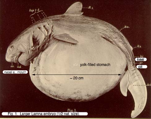
| Fig. 1. Larger Lamna-embryo. (1/2 nat. size.) [TL = 55.3 cm] |
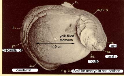
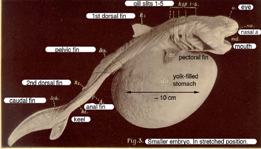
Fig. 2. Smaller embryo. In natural position. (Ca. 1/2 nat. size.) [TL = 42.8 cm] | Fig. 3. Smaller embryo. In stretched position. (Ca. 1/2 nat. size.) |
| A = anal fin; Ba = pelvic fins; Br = pectoral fins; Do = yolk stomach; Gr = precaudal pit; ki = caudal keel; ksp1-5 = 5 gill slits; md = mouth; na = nasal aperture; o = eyes; R1 = 1st dorsal fin; R2 = 2nd dorsal fin; Sch = caudal fin. | ||
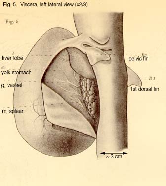
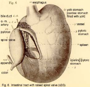
| Fig. 4. Viscera, right lateral view (2/3 nat. size). | Fig. 5. Viscera, left lateral view (2/3 nat. size). | Fig. 6. Intestinal tract with raised spiral valve (2/3 nat. size). |
| a.o.m. = omphalo-mesenteric artery; app = appendix; Ba = pelvic fins; Br = pectoral fins; Do = yolk stomach [cardiac stomach filled with yolk]; d. ch = bile duct; e = colon; l = right or left liver lobe; m = spleen; mp = pyloric stomach opening; oe = esophagus; p = pyloric stomach; py = pylorus; R1 = first dorsal fin; s = valvular intestine (spiral valve). | ||
| Fig. 7. "Had to be deleted." according to Lohberger (1910). | ||
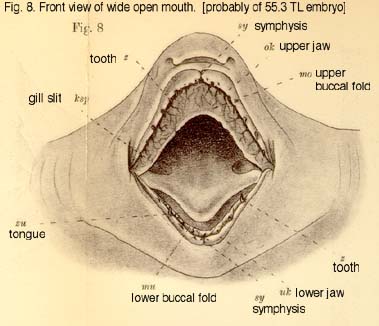
| Fig. 8. Front view of
mouth, wide open. |
Ksp = gill slit; mo = upper buccal fold [maxillary valve]; mu = lower buccal fold; ok = upper jaw; sy = sympheses; uk = lower jaw; z = teeth; zu = tongue. | |
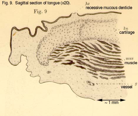
| Fig. 9. Sagittal section of tongue (x20). | g = vessel; kn = cartilage; hz = recessive mucous denticle; mus = muscle. | |
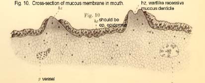
| Fig. 10. Section of mucous membrane in mouth. | ep = epidermis; g = vessel; hz = wartlike recessive mucous denticles. | |
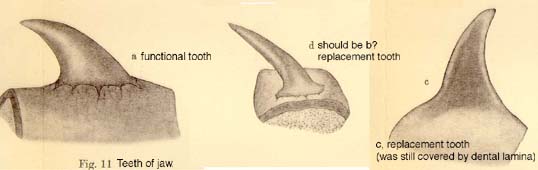
| Fig. 11. Teeth of jaw [rather than skin]. | a = fully formed tooth [functional]; b = replacement tooth [mis-labled "d"]; c = replacement tooth (still embedded in dental laminae). | |
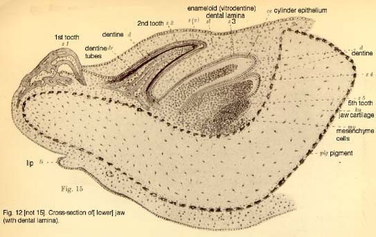
| Fig. 12. Cross-section through jaw (with dental lamina). [lower jaw, probably of smaller embryo]. | ||
| ce = cylinder ephithelium; d = dentine; dr = dentine tubes;
kn = jaw cartilage; li = lip formation; my = mesenchyme cells; pig = pigment;
s (v) = enameloid; z1-z5 = teeth; zl = dental lamina. [z1 has an extremely curved crown unlike the replacement teeth still in the dental lamina. R. Purdy commented that this tooth is similar in form to those found in stem chondrichthyans and that this curvature may be the expression of a primitive character which is lost in later generations.] |
||
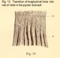
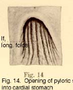
| Fig. 13. Transition of longitudinal folds into net of folds in the pyloric stomach. | Fig. 14. Opening of pyloric stomach from cardiac stomach. | lf = longitudinal folds; fn = net of folds? |
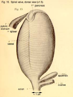
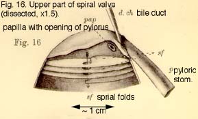
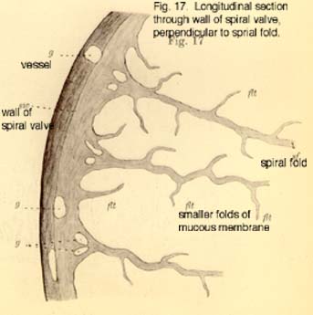
| Fig. 15. Spiral valve, dorsal view. (x1.5). | Fig. 16. Upper portion of spiral valve. (opened, x1.5). | Fig. 17. Cross-section through wall of spiral valve, perpendicular to spiral folds. |
| app = appendix, d.ch = bile duct; d = colon; flt = mucous fold?; g = vessel; m = spleen; of = openings between spirals?; ov = ovaries; p = pyloric stomach; pa = pancreas; pap = papilla with pyloric valve; sf = spiral fold; sw = wall of valvular intestine [spiral valve]. | ||
| Fig. 18. Cross-section of appendix (x50). | Fig. 19. Longitudinal section of appendix (x50). | Fig. 20. Longitudinal section of appendix near surface (x55). |
| Fig. 21. Cross-section of appendix near opening. | be = cup cells?; cy = cylinder cells; dr = glandular tubes; g = vessels; gg = larger vessels sitting on mucous folds?; mus = muscle layer; sh = mucous membrane; shf = mucous folds; *) part of gland, free of mucous folds and [blood] vessels. | |
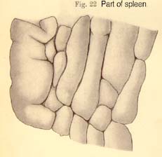
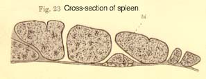
| Fig. 22. Part of spleen. | Fig. 23. Cross-section of spleen. | bi = epidermis of connective tissue. |
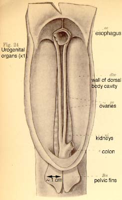
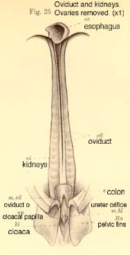
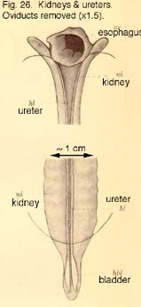
| Fig. 24. Urogenital organs (nat. size). | Fig. 25. Oviduct and kidneys. Ovaries removed. (nat. size). | Fig. 26. Kidneys and ureters. Oviduct removed (x1.5). |
| Ba = pelvic fins; dlw = dorsal body cavity wall; e = colon; eil = oviduct; hbl = bladder; hl = ureter; kl = cloaca, m.eil = orifice of oviduct; m.hl = opening of ureter; ni = kidneys; up = papilla of ureter; oe = esophagus; ov = ovaries. | ||
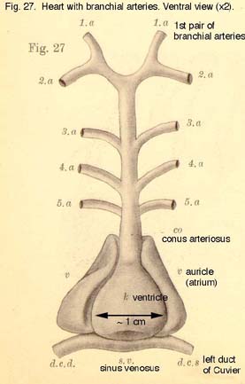
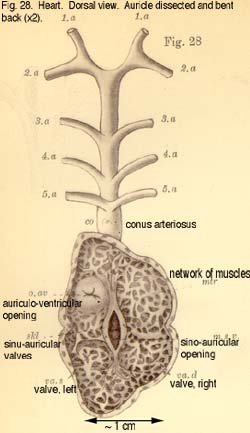
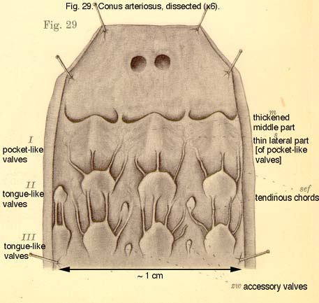
| Fig. 27. Heart with branchial arteries. Ventral view (x2). | Fig. 28. Heart. Dorsal view. The auricle is dissected and bent backwards (x2). | Fig. 29. Conus arteriosus, dissected. (x6). |
| 1a-5a = 5 branchial arteries; co = conus arteriosus; d.c.d = duct of Cuvier, right; d.c.s = duct of Cuvier, left; k = ventricle; ka = opening of the 5 branchial arteries [not used in any figure?]; m = thickened middle part [of pocket-like valves]; m.s.v = sinu-auricular opening; mtr = network of muscles; o.av = auriculoventricular opening; s = thinner lateral part [of pocket-like valves]; sef = tendinous chords; skl = sinu-auricular valves; s.v = sinus venosus; v = auricle (atrium); va.d = right sinu-auricular valve; va.s = left sinu-auricualar valve; zw = accessory valves; I-III = transverse rows of valves; I = pocket-like valves; II, III = tongue-like valves. | ||
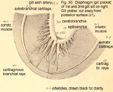
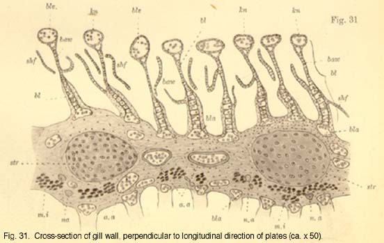
| Fig. 30. Diaphragm [gill pocket] of 1st and 2nd gill slit on right hand side (nat. size). | Fig. 31. Cross-section of gill wall. | |
| baw = Balkenwerk?; bl = gill filaments; bla = afferent branchial arterioles; blv = efferent branchial arterioles; ep = epibranchial with cartilaginous branchial rays; kb. a = branchial arch artery. branchial afferent arteries, arterioles are shown in black for clarity; ker = ceratobranchial with cartilaginous branchial rays; kh = extra-branchial cartilage originating at hypo-branchial; kn = button-like end of gill filament [containing efferent branchial arteriole] kp = extra-branchial cartilage originating at pharyngo-branchial; m. add = adductor muscle; m.c = constrictor muscle; m. i = interbranchial muscle; n.a = nerve branches?; shf = small mucous membrane folds, dissected irregularly. | ||
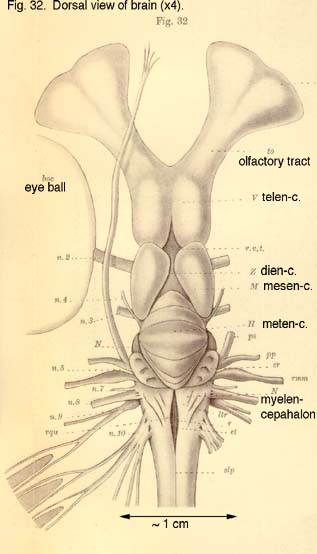
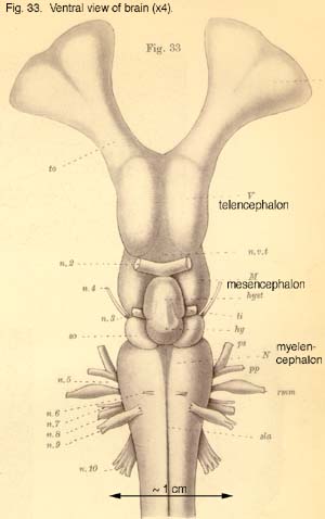
| Fig. 32. Dorsal view of brain (x4). | Fig. 33. Ventral view of brain (x4). | |
| V = telencephalon; Z = diencephalon; M = mesencephalon [optic
lobes]; H = metencephalon [cerebellum]; N = myelencephalon [medulla oblangata
& restiform bodies]. Cranial nerves: [n.1 = olfactory, not shown]; n.2 = optic; n.3 = oculomotor; n.4 = trochlear; n.5 = trigeminal; n.6 = abducens; n.7 = facial; n.8 = auditory; n.9 = glossopharyngeal; n.10 = vagus. boc = eyeball; cr = restiform bodies; et = eminentiae teretes; hy = pituitary gland; hyst = epithalamous; li = infundibulum [& asssociated structures?]; lo = olfactory bulb; ltr = lobi trigemini; p.p = ophthalmicus profundus branch; p.s = ophthalmicus superficialis branch; rmm = maxillary branch [of trigeminal]; r.v.t = regio ventriculi tertii; sla = sulcus longitudinalis anterior; slp = sulcus longitudinalis posterior; sv = vascular sac; to = olfactory tract. |
||
| Fig. 34. Median, perpendicular cross-section of vertebrae (x6). | a = outer zone; cd = centrum; fc = notochord; h = haemal arch; i = inner zone; n = neural arch; st = calcified rays [spokes]. | |
Created October 1999; updated April 2007. Back to previous page