Photo List II for Morro Bay Great White Shark
Generated by C.R. Davis and D.B. Harris


(Was found in anterior end of liver and later identified to have come from Myliobatis californica.
|
Photo List II for Morro Bay Great White Shark Generated by C.R. Davis and D.B. Harris |
||
|---|---|---|
|
||
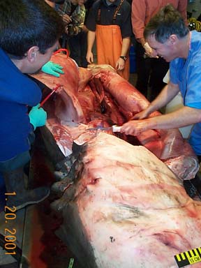
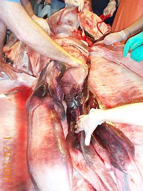
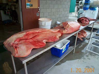
| Back to Photo List I | Back to Photo List I | Photo #59. Dissection. |
|
. |
|
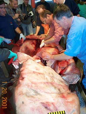
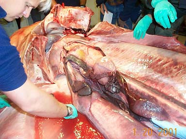
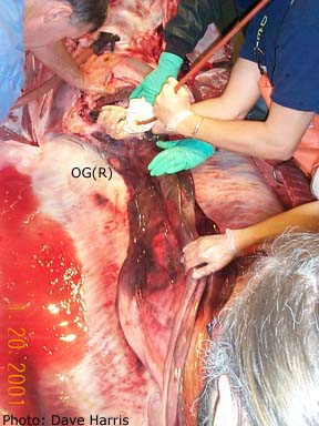
| Photo #60. Removing the liver (Dave Casper and Dave Ebert) | Photo #64. Removing of the liver. | Photo #66. Upper gastrointestinal tract, in situ |
|
|
|
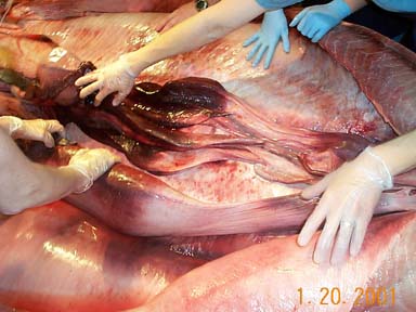
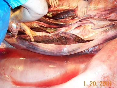
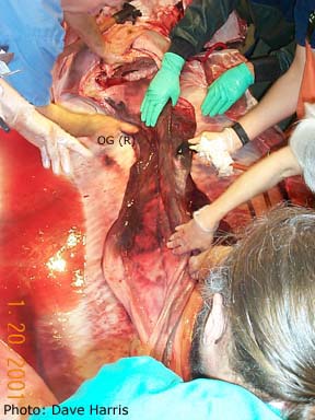
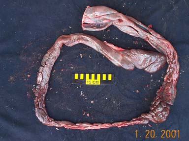
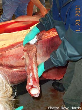
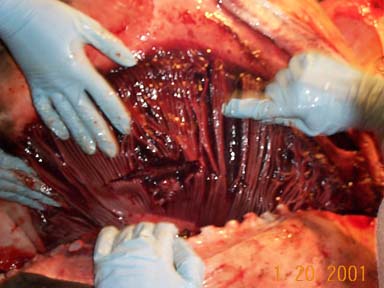
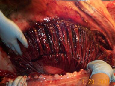
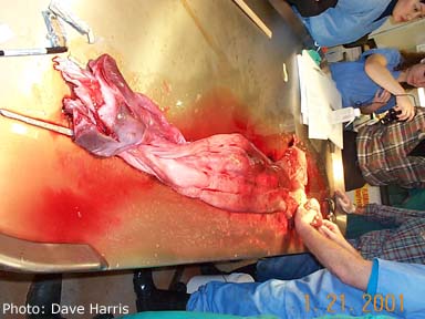
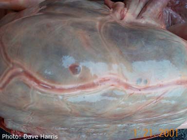
| Photo #69. Gastrointestinal tract, in situ. Valvular intestine in near bottom, right center; long, multilobular pancreas and stomach near center. | Photo #70. Valvular intestine and colon near bottom; female reproductive tract is above gastrointestinal tract. Rectal gland is located just dorsal to the colon. | Photo #71. Gastrointestinal tract and reproductive tract. |
|
|
|
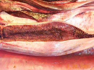
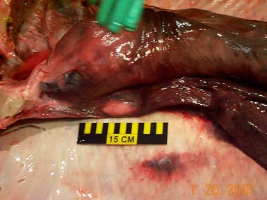
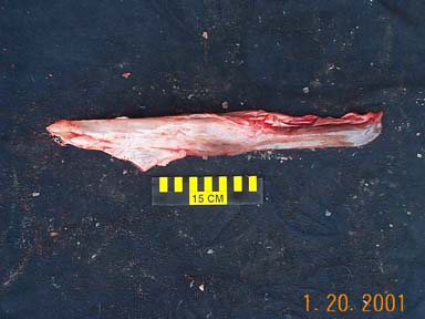
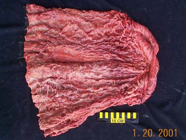
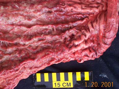
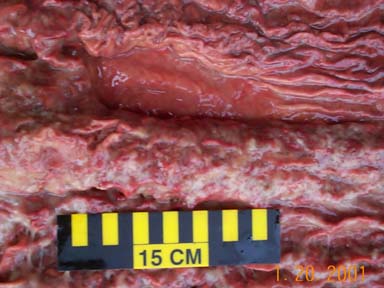
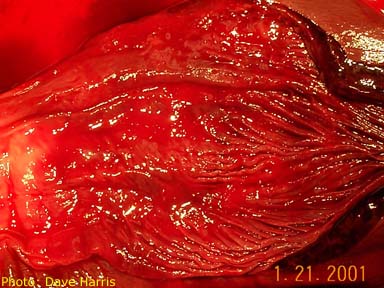
| Photo #73. Valvular intestine, note rectal gland dorsal to colon, at right of photo. | Photo #74. Valvular intestine. | Blank |
|
|
|
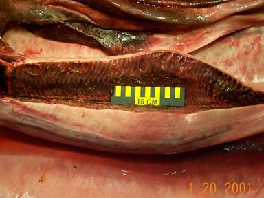
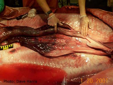
| Photo #76. Valvular intestine. | Photo # 77. Reproductive tract, in situ. | Photo # 78. Reproductive tract, in situ. |
|
|
|
|
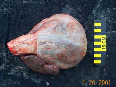
| Photo # 79. Female reproductive tract, in situ; shell gland immediately above marker. | Photo # 80. Reproductive tract, in situ. | Photo # 81. Liver. |
|
|
|
| Photo #84. Pancreas. | Photo # 85. Spleen. | Photo # 87.Vertebral column. |
|
|
|
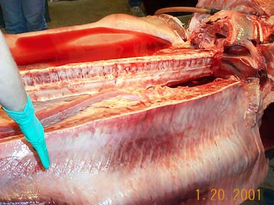
| Photo #88. Vertebral colum. | Photo # 89. Vertebral column | Photo # 90 Gastric mucosa. Note region lacking rugal folds near left-center. |
|
|
|
| Photo #91. Rugal folds of gastric mucosa, close-up. (Does not include smooth region). | Photo #92. Region of gastric mucosa lacking rugal folds, close-up. | Photo #93. Vertebral centrum. |
|
|
|
| Photo #94. Ampullae of Lorenzini, ventral snout. | Photo # 95. Sting ray spine. (Was found in anterior end of liver and later identified to have come from Myliobatis californica. |
Photo # 96. Gill filaments. |
|
|
|
| Photo #97. Gill filaments. | Photo #98. Heart ventricle, conus arteriosus and atrium. | Photo #99. Epigonal organ. |
|
|
|
| Photo #100. Female reproductive tract. | Photo #101. Epigonal organ. | Photo #102. Epigonal organ. |
|
|
|
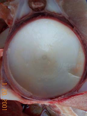
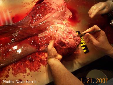
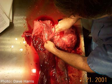
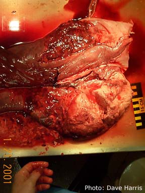
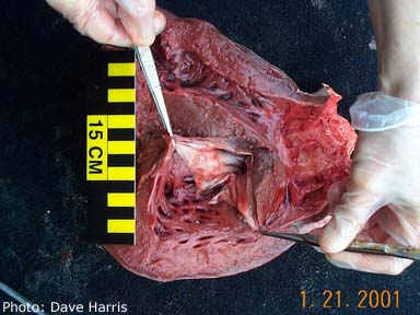
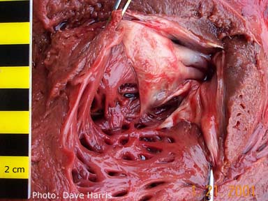
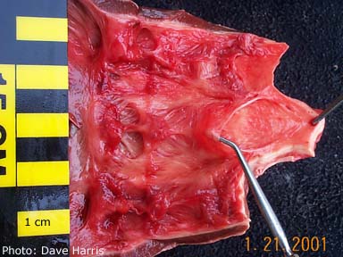
| Photo #103. Ventricle of heart: view of atrioventricular (mitral) valve. | Photo #104. Ventricle of heart: view of atrioventricular (mitral) valve, close-up. | Photo #105. Close-up of semilunar valvee in conus arteriosus, with probe in one of the membranous valve leaflets. |
|
|
|
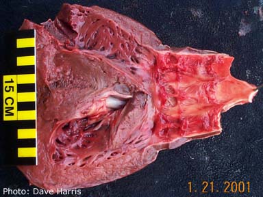
| Photo #106. Ventricle of heart opened to show base of conus arteriosus and 2 sets of semilunar valves. | Photo #107. Small fluid-filled subepicardial cysts. | Photo #112. Red, congested uterus. |
Created April 2001; updated July 2001. Back to previous page